Scientists for the first time captured the process of embryo formation at an early stage of development (6 photos + 1 video)
Australian researchers from the University of Queensland made a unique discovery - for the first time they managed to film the process of embryo formation at the earliest stages. Scientists hope this will help unlock medical mysteries and better understand how birth defects occur in humans. 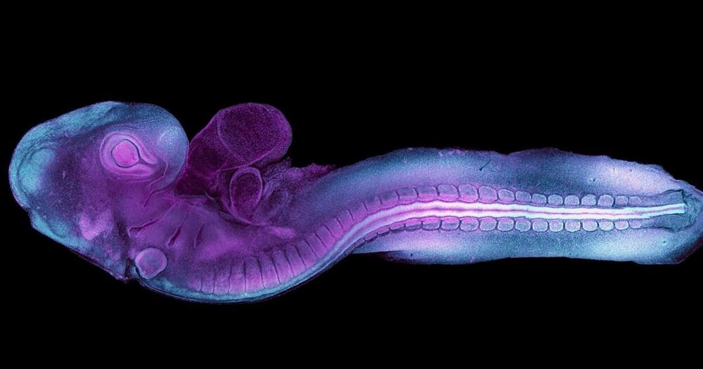
“Bird embryos are an excellent model of human development, especially in the early stages of growth,” said lead author Dr Melanie White. The development of many major organs, including the heart and the neural tube (which later forms the brain and spinal cord), is very similar in birds and humans, she said.
For the first time in history, scientists have recorded real-time video of an embryo forming a neural tube at an early stage of development, which will later become the brain and spinal cord.
For their observations, the scientists used a genetically modified quail embryo that produced a reflective fluorescent protein.
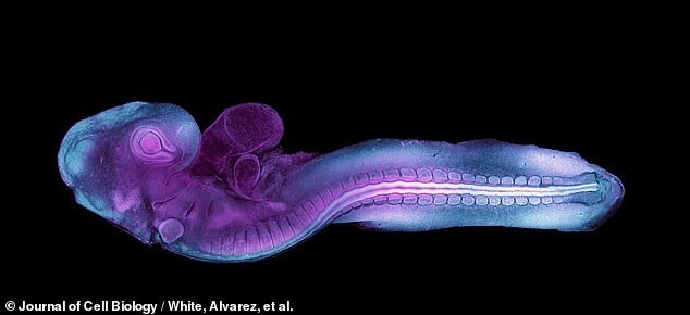
An innovative technique using fluorescent protein has illuminated this tiny embryo.
This allowed them to look in detail at how embryonic cells crawl along a protein support structure, organizing themselves into the earliest form of the heart and the first phase of the spine and brain.
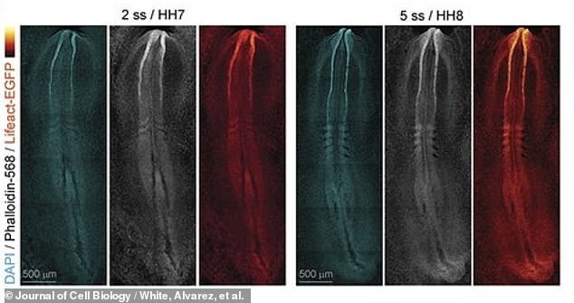
Researchers hope these new videos can help modern medicine understand what birth defects are and how to correct them. The picture shows footage of the early formation of the spine and brain of the embryo.
“We saw the cells extending their projections through the open neural tube to communicate with the opposite side,” Dr. White said. “The more protrusions the cells formed, the faster the tube “zipped up.”
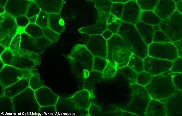
The glow of fluorescent proteins reveals the embryo's early scaffolding, called the "actin cytoskeleton." It gives cells shape and helps them move.
It is this process that often "fails or becomes disrupted" during the fourth week of human development, leading to congenital malformations of the brain or spine, both hereditary and caused by environmental factors.
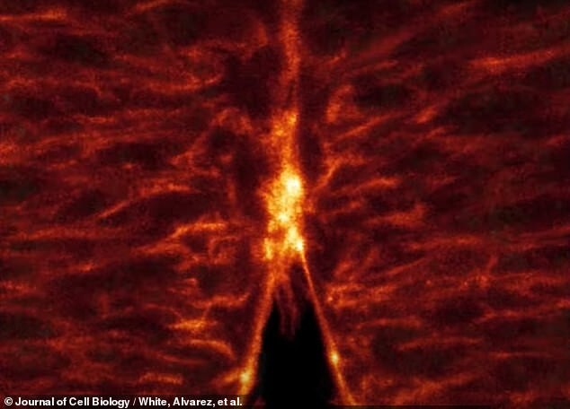
The image shows the "zipper" movement as the embryo's neural tube forms as the arm-like projections of each cell - lamellipodia and filopodia - cling to each other.
“Our goal is to find proteins or genes that can be targeted or used in the future to screen for birth defects,” says Dr. White.
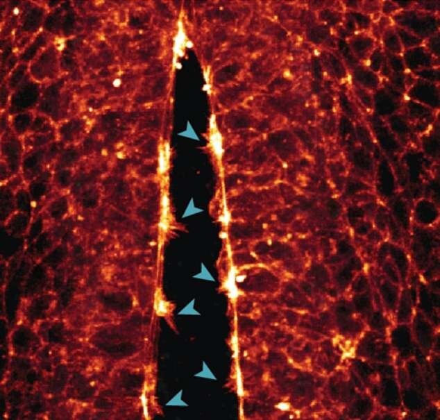
The researchers were able to detect the formation of hand-like protrusions on individual cells, which help the cells crawl along the protein supports of the cytoskeleton to the desired location. In the image of the hand, the cells join together to close the walls of the neural tube.
Scientists hope the new quail embryo model will allow them to study in real time which early mistakes in embryonic cells lead to birth defects, to help improve treatments for humans.


