25 unique photographs that show in detail the process of conception and intrauterine development of a child (22 photos)
The birth of a new life and the birth of a baby is an amazing phenomenon that can be called a miracle! Two loving people, a man and a woman, create the fruit of their love - their own likeness, a little person, whose appearance further strengthens and strengthens the feelings of the new parents. And this is wonderful! 
The creation of a child is a mystery. But restless scientists, striving to study everything in the world, were able to track how this mystery occurs, and how further, after conception, the fetus, the future little person, develops. Thus, the Swedish scientist and photographer Lennart Nilsson managed to do this back in the 50s of the last century. Of course, it was not easy to do back then. The scientist tried to use different techniques, including macro lenses. He achieved the best results by attaching a micro-illuminator and a micro-camera to the end of a cystoscope tube. Without causing any harm to the future baby, Lennart photographed all stages of its development, from conception to the stage when the fetus is ready to be born.
This painstaking and incredibly complex work took Lennart Nilsson a whole decade. By 1965, he finally got excellent quality pictures. And in the same year, they were published in the then popular American magazine LIFE. The name of the scientist-photographer immediately became known throughout the world. Having seen his work, you will agree that he is worthy of such fame!
The moment of movement of the sperm to the egg through the fallopian tube 
What a beauty - the egg! 
The moment when a sperm meets an egg, resulting in a new life 
In 1965, Lennart Nilsson published a wonderful book called "A Child is Born", with a large number of colorful illustrations. On its pages one could see photographs of embryos in development, as well as comments and explanations from doctors, their recommendations for carrying a healthy fetus. Later, several more of his books were published, which contained mainly photographs on the same subject. A frame that captured the moment of conception, when the sperm broke through the egg shell and merged with it 
After seven days, you can see the blastocyst emerging from the fertilized cell, which is a ball of embryonic cells 
The embryo moves into the uterus, where it continues to develop.
On the eighth day 
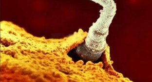
The moment of embryo attachment to the mucous surface of the uterus. On the twenty-second day
The embryo is actively developing: the gray zone will be the brain of the fetus.
On the twenty-fourth day 
On the 24th day of the embryo's life, it will be marked by the appearance of a primitive heart, which already has 2 chambers (ventricles). This organ plays a major role in further development, as the heart ensures blood circulation, distributing oxygen and nutrition to the entire embryo, including the small organs that are beginning to develop.
At four weeks 
At four weeks, the embryo is approaching seven millimeters in size. It is already forming a body with a head and tail.
At five weeks 
At five weeks, the size of the future baby is nine millimeters. During this period, it begins to form a semblance of a face with holes for the eyes, nose and mouth.
At six weeks 
Its size increases to twenty millimeters.
At the age of seven weeks 
During this period, the body of the future baby continues to grow. And if earlier the embryo resembled the embryo of a primitive mammal, now it is already clear that it will be a little man. The cranial bones are not yet dense enough, and the brain can be seen through them. Moreover, in the forehead area, two bubble-like convex structures are clearly visible, from which the brain is formed.
At the age of eight weeks 
At this stage, the future baby weighs about eight grams and is approximately thirty millimeters long. Not only is its heart well developed, but its head, legs, and arms are also well developed.
At the age of nine weeks 
At this stage of its development, the fetus becomes even more human-like. In places where the cranial bones grow together, many blood vessels appear.
At twelve to thirteen weeks 
The photo shows that on the left side of the small fetus there is a yolk sac that provides the embryo with nutrition until the full placenta is formed.
At thirteen weeks 
This is what the embryo looks like at this time. It is well protected.
At the age of sixteen weeks 
Through the thin, delicate skin of the future baby, we see blood vessels. He is already beginning to explore his body and what surrounds him with his hands.
At the age of eighteen weeks 
At this age, the fetus can reach fourteen centimeters in length. At this stage, he can already hear sounds from the outside.
At twenty weeks old 
His body and head begin to be covered with hair.
At twenty-three weeks 
At twenty-six weeks 
The length of the fetus at this age reaches thirty centimeters.
At thirty-six weeks 
A few more weeks and the baby will be born.
Lennart Nilsson has made a huge contribution to science! By combining his amazing creativity and traditional medicine, he has significantly expanded the horizons of photography.