How a child develops: the photographer showed the magical process of the birth of a new life (23 photos)
The birth of a person is a real miracle, which even modern science never ceases to admire. Scientists around the world are constantly studying this process, trying to learn as many new details about it as possible. Fortunately, innovative technologies make this possible. 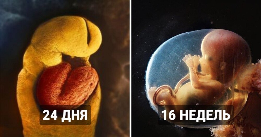
However, back in the 1950s, Swedish photographer and part-time scientist Lennart Nilsson did the truly impossible. He managed to film the evolution of the embryo from conception to birth and obtain informative and extremely interesting images. How did he manage to do this, even in such a low-tech period? Of course, Lennart did not succeed all at once. He experimented with different techniques and used macro lenses. However, he was able to obtain the best result by adapting a microcamera and a microilluminator to the end of the cystoscope tube. Without disturbing the process of fetal development, the photographer captured all the stages that occur in a woman’s womb during pregnancy.
The photographer spent ten years trying to get the best possible pictures. In 1965, they were published by LIFE magazine (very popular in the USA at that time), thanks to which Lennart Nilsson gained tremendous popularity. Nilsson has created something truly wonderful. Thanks to him, people for the first time were able to see with their own eyes the origin and early development of human life.
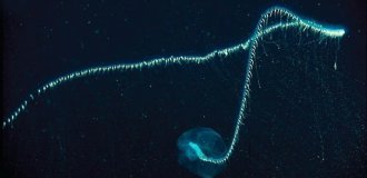
The sperm moves down the fallopian tube to the egg 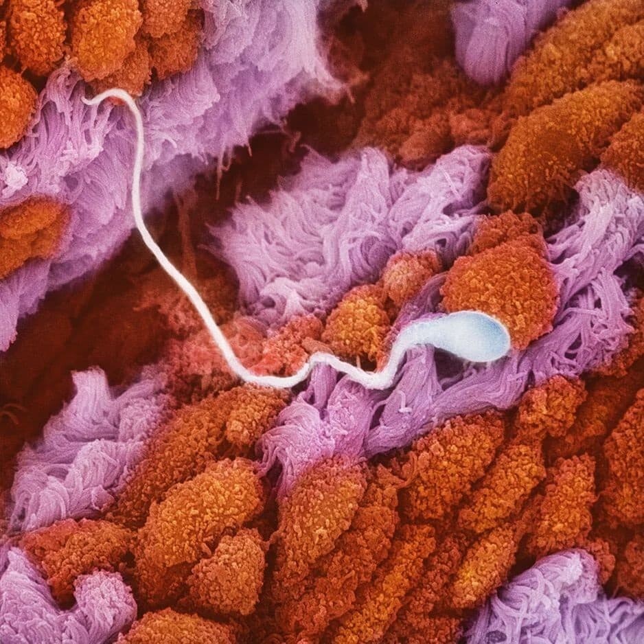
This is what an egg looks like 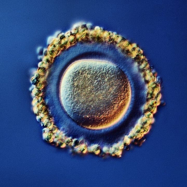
Fateful meeting 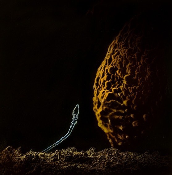
In 1965, Lennart Nilsson published an illustrated book called A Child Is Born, which included not only photographs of developing embryos, but also texts from doctors, as well as advice on prenatal care. Subsequently, the photographer published several more books. They already focused primarily on photography.
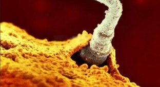
The sperm breaks through the membrane of the egg and flows into it 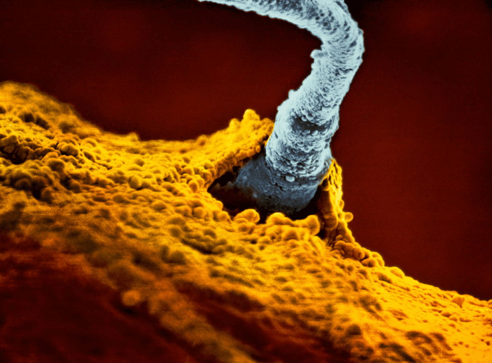
A week later, a blastocyst appears - a ball of embryonic cells arising from a fertilized egg. 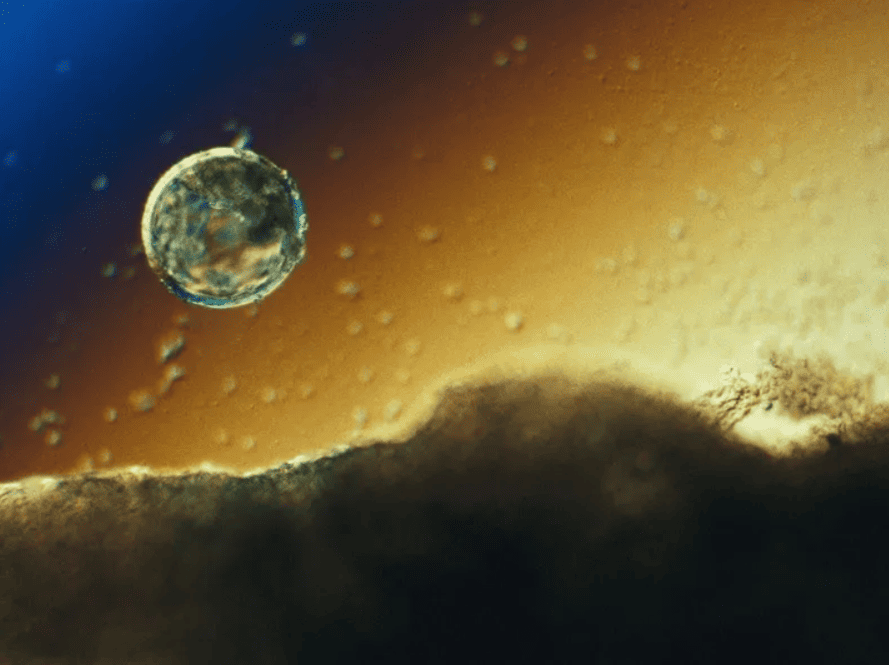
The embryo moves into the uterus and is fixed.
8 days 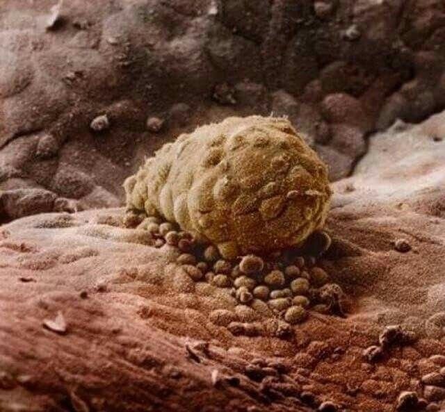
The embryo attaches to the lining of the uterus.
22 days 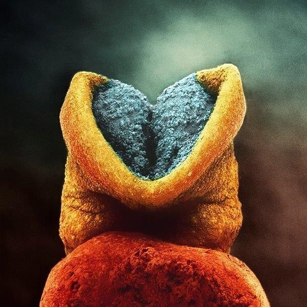
The development process begins. The gray area will become the child's brain.
24 days 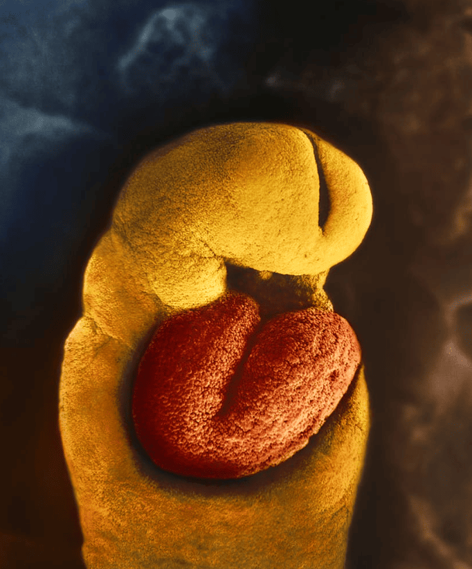
The embryo develops a primitive heart. It already has two chambers (ventricles). The heart plays a major role in the development of the embryo, because it is responsible for the circulation of blood, which distributes nutrients and oxygen to all the small developing organs.
4 weeks 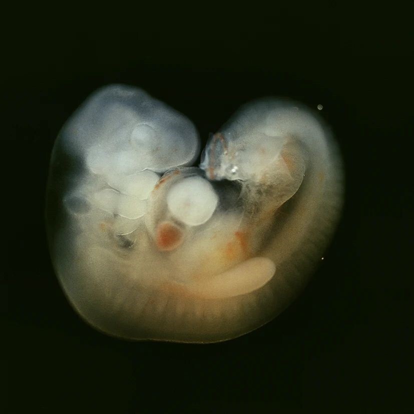
The dimensions of the embryo are about 7 millimeters. Now he has a body with a head and a tail.
5 weeks 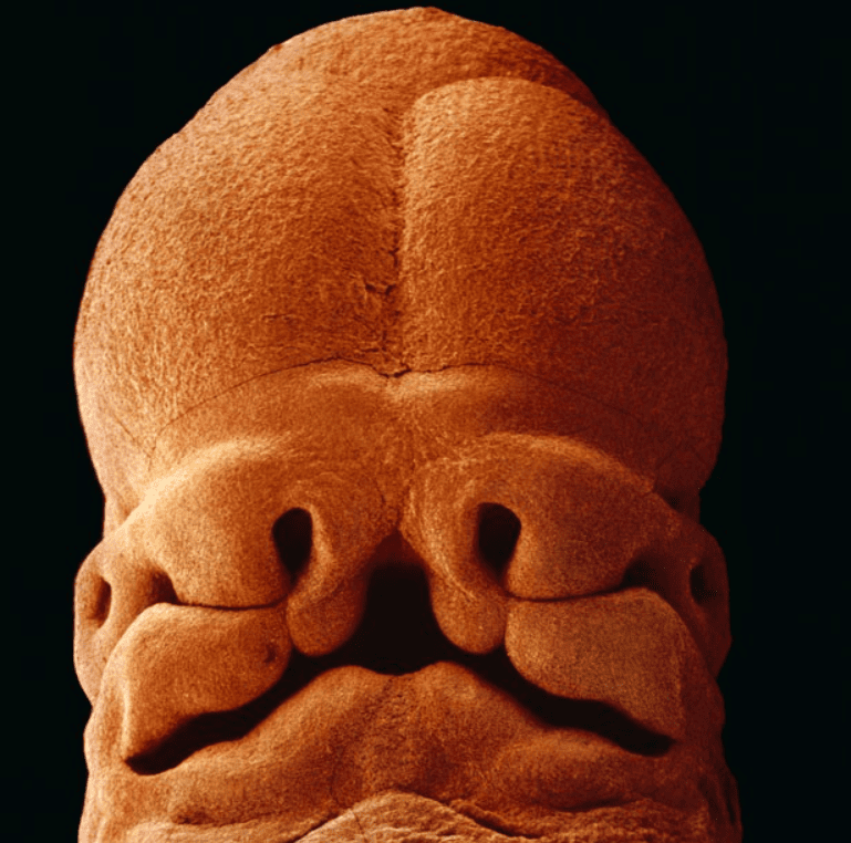
At five weeks, the embryo grows to 9 millimeters. He develops something like a face, in which openings for the future mouth, nose and eyes are already recognizable.
6 weeks 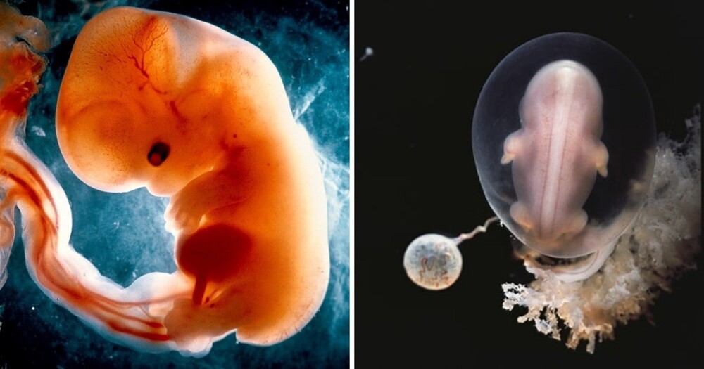
The dimensions of the embryo are about 20 millimeters.
7 weeks 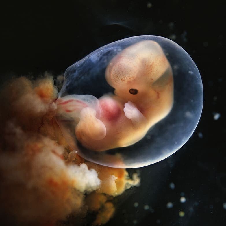
The body continues to grow. What once resembled the embryo of any primitive mammal is now beginning to look like a miniature human being. The bones of the skull are not yet very dense, so the brain is visible through thin cartilage. In the forehead area, two large bubble structures are visible that will soon become the brain.
8 weeks 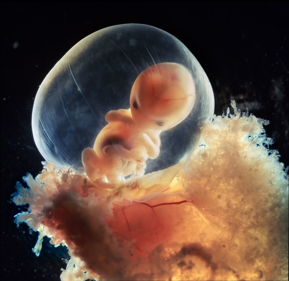
The fruit at this stage is about 30 millimeters long and weighs about 8 grams. The heart, arms, head and legs are developed.
9 weeks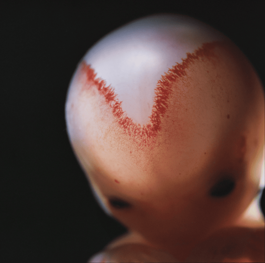
The fetus becomes more and more human-like. A large number of blood vessels are formed at the fusion sites of the skull bones.
12-13 weeks 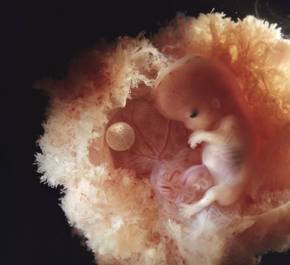
To the left of the fetus is the yolk sac, which provides nutrition to the embryo until the placenta is fully formed.
13 weeks 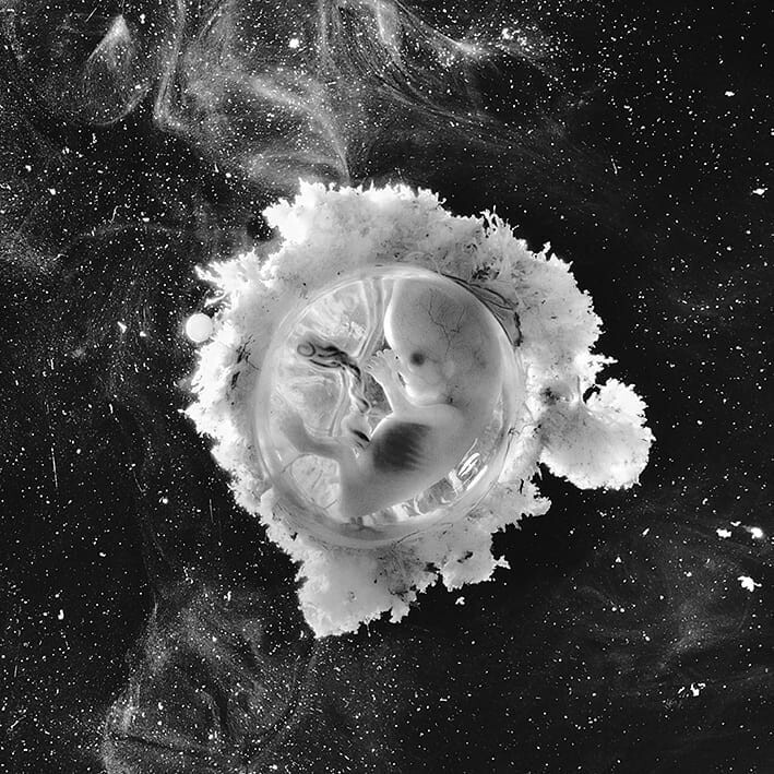
The fast-growing embryo is reliably protected in the mother's womb.
16 weeks 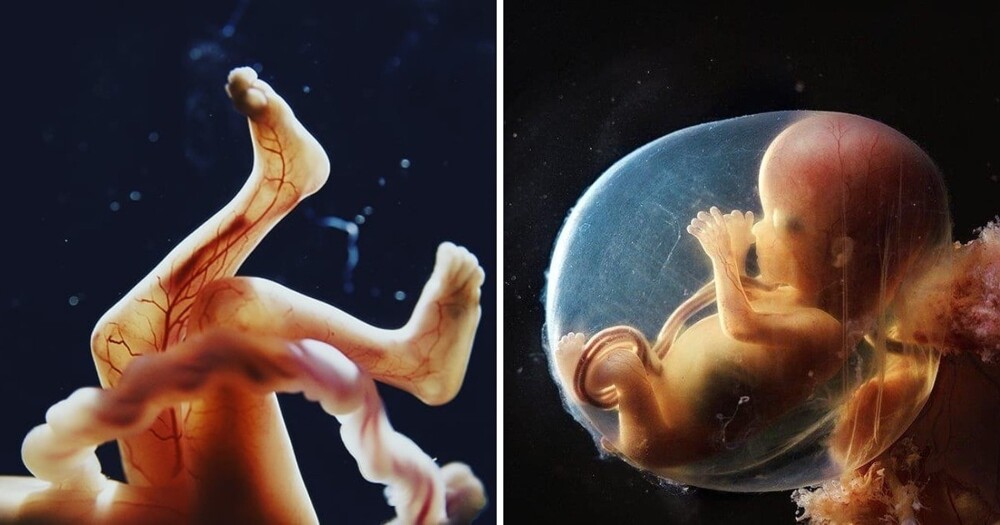
A network of blood vessels is visible through the thin skin. The fetus begins to use its hands to explore its body and environment.
18 weeks 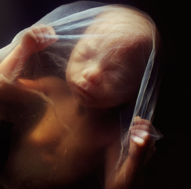
The fruit measures approximately 14 centimeters. Now he can perceive sounds from the outside world
20 weeks 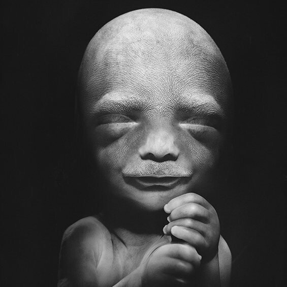
Hair begins to cover the head and body.
23 weeks 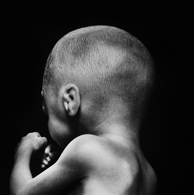
26 weeks 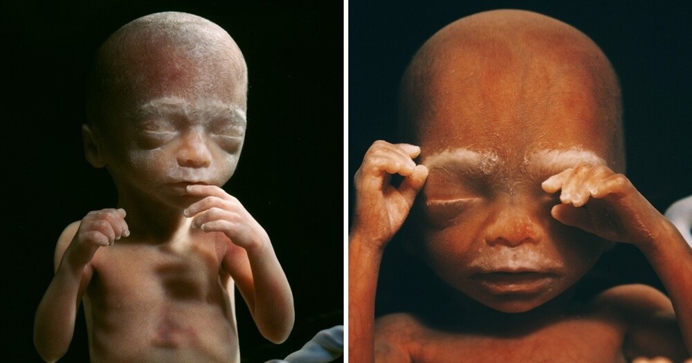
At this stage of development, the size of the fetus is about 30 centimeters.
36 weeks 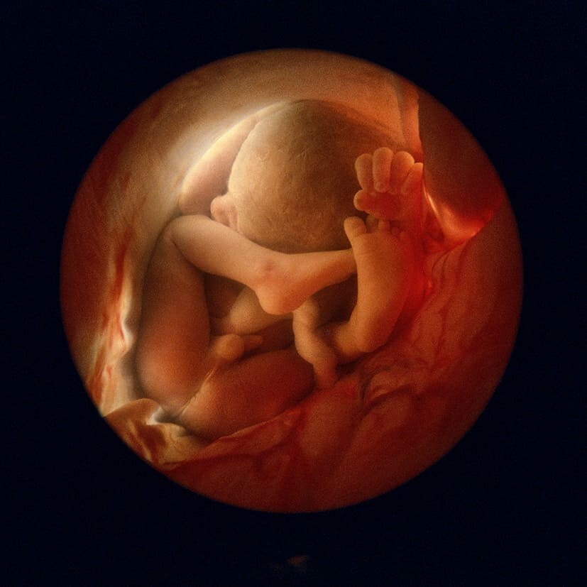
Very soon the baby will be ready to be born.
Lennart Nilsson made great contributions to science. He was able to combine medicine and creativity, which in the future made it possible to open new horizons in photography.