Italian scientists recreated the face of a pregnant Egyptian mummy (12 photos + 1 video)
Last year, Polish scientists studied an ancient Egyptian mummy known as the "mysterious lady", and found in her stomach signs of germ. The researchers found that the woman died about 2000 years back at 28 weeks pregnant between the ages of 20 and 30. Now on based on her remains, Italian experts created two images, which show what she might have looked like when she was alive in the first century BC ad. 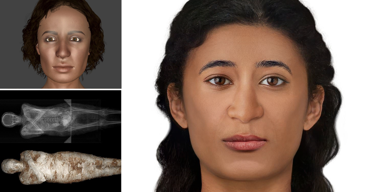
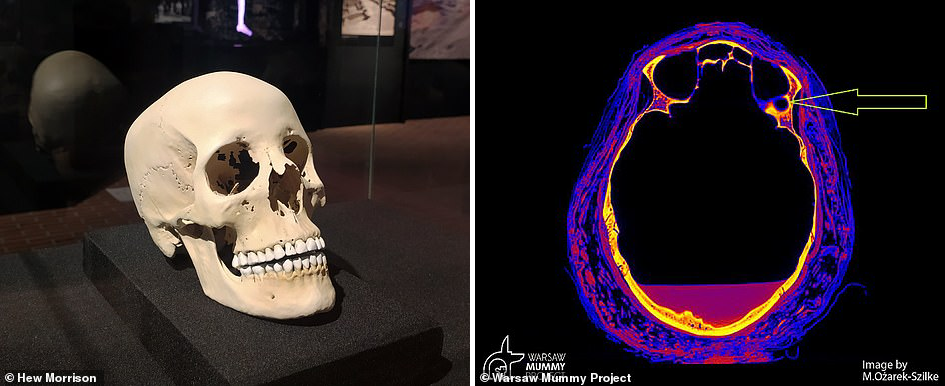
“Our bones, and in particular the skull, provide us with much face information,” says Chantal Milani, an Italian judicial anthropologist and participant in the Warsaw Mummy project. - Although it is not possible called an accurate portrait, the skull, like many parts of anatomy, is unique and demonstrates a set of shapes and proportions, which eventually appear in face."
“The face covering the bone structure follows different anatomical rules, so for its reconstruction can be used standard procedures, for example, to determine the shape of the nose, added she is. – The most important element is the reconstruction of the thickness of the soft tissues at numerous points on the surface of the facial bones. For this, we have statistics for different populations around the world." 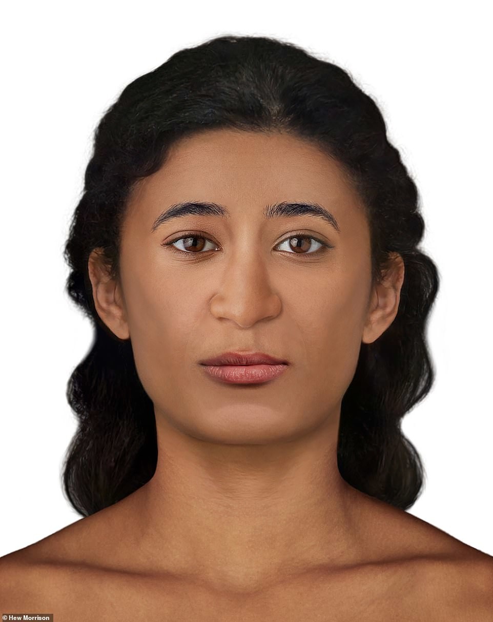
The “Mysterious Lady” was found in the early 1800s during the Tsarist tombs in Thebes and belonged to the Theban elite. Mummy dated the first century BC, the time when Cleopatra was queen, and Thebes was a lively city. The mummy was transported from Egypt to Warsaw in December 1826, during the period of the most important discoveries in the Egyptian Valley kings. Initially, it was believed that this was the mummy of the priest Khor Dzhehuti, but in 2016, scientists found that carefully wrapped cloth the embalmed body belongs to a woman. This is the first mummy in history, containing fruit and is currently on display at the National museum in Warsaw. 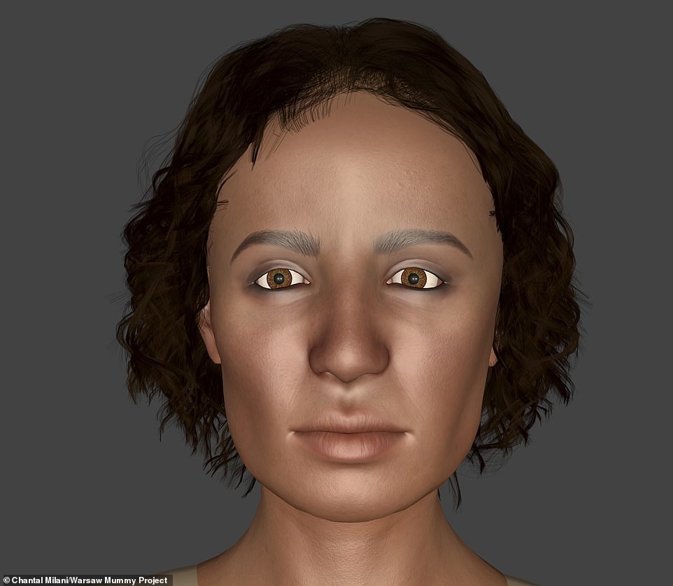
Two forensic experts, along with researchers from University of Warsaw decided to "revive" the mummy using 2D and 3D -technology for the reconstruction of her face. Medical Examiner Hugh Morrison said: “Mostly facial reconstruction is applied in forensics to help recognize the body when more common means of identification such as fingerprints or DNA analysis yielded no results.
"Reconstruction of a person's face from his skull is often considered last resort in an attempt to identify him. Morrison continued. — It can also be used in an archaeological and historical context, to show how ancient people or prominent personalities of the past could look like in real life. In a historical context, this process helps figuratively bring the deceased back to life, and also develops respect and sensitivity to deceased who are either the subject of research or exhibited in museums. 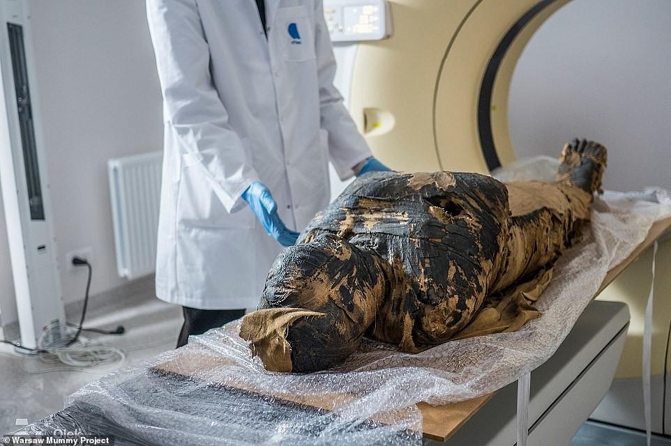
Dr. Wojciech Eismond, archaeologist from the Polish Academy of Sciences, added: “For many people, ancient Egyptian mummies represent curiosity, and some people tend to forget that they were once living people who had their own lives, loves and tragedies. It can be said that forensic experts provide faces to scientific data, therefore, the person is no longer an anonymous "curiosity in the window". In addition, it was very important for the ancient Egyptians to preserve their appearance. the deceased to help his soul and personality survive. So, from one on the other hand, we are, so to speak, "rehumanizing" scientific data, and on the other – we fulfill the desire of these people not to be anonymous and forgotten.”
The reconstruction of the mummy's face can be seen at the exhibition in the Silesian Museum in Katowice from November 3rd. 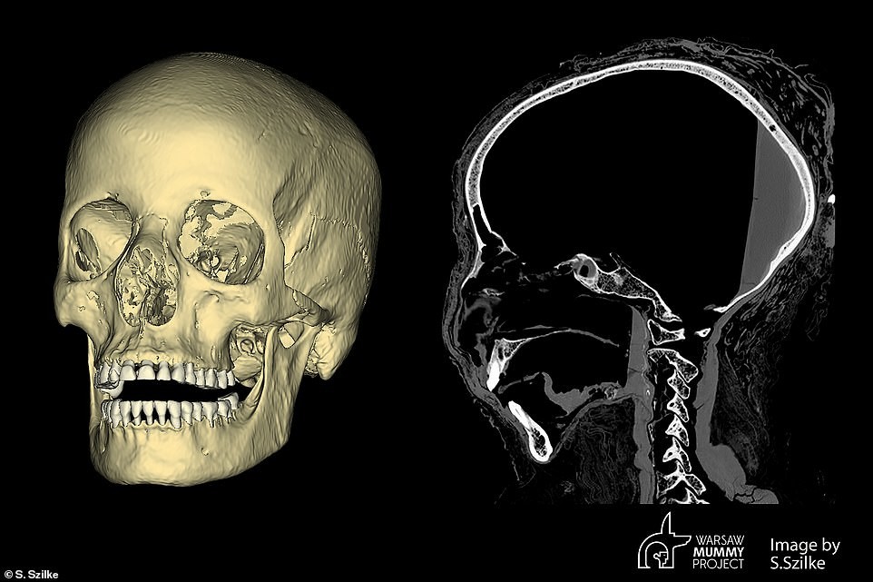

As for the fetus, it is located in the lower part of the small pelvis and partly in the lower part of the large pelvis of the mother and was mummified together with her. Surrounding tissue obscures fetus on CT images uterus, so scientists were only able to measure the circumference of his head. She is was 24.9 centimeters, which suggested that the fetus was between the 26th and 30th week of life. The uterus and the fetus itself were not taken out, as in the case of internal organs, including the heart, lungs, liver and intestine with stomach.
Project experts"Warsaw Mummy" can't tell why the fetus was not extracted and mummified separately, as was the case with stillborn. “Perhaps it was believed that he was still part of the body of his mother, because he was not yet born, ”the scientists said at that time time. It turns out that, according to the beliefs of the ancient Egyptians, the unborn the child could live the afterlife only if went to the next world with his mother. 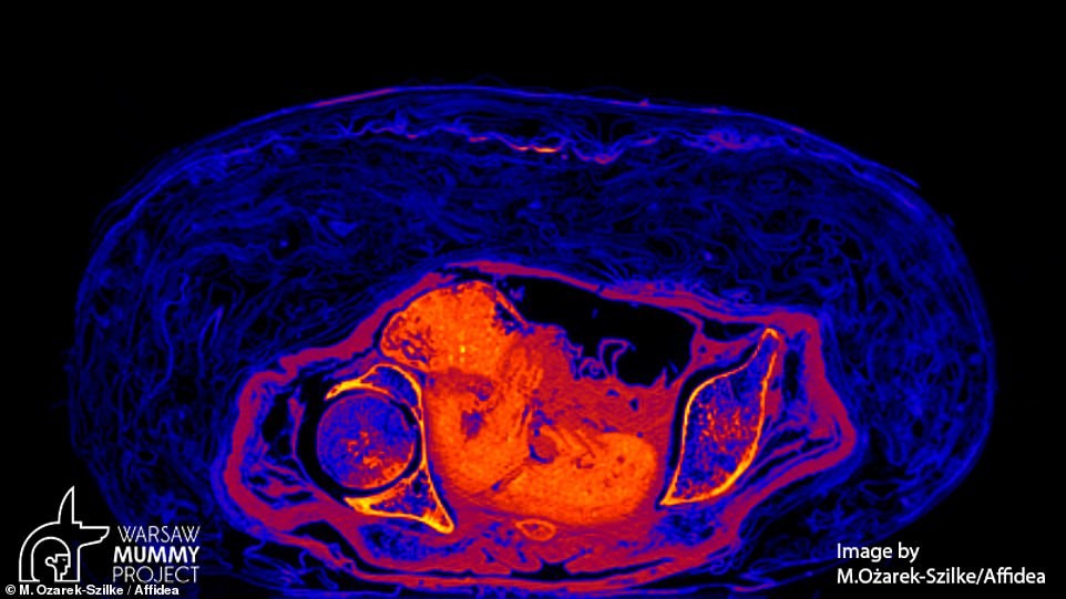
In 2021, researchers from the Polish Academy of Sciences published article about the first pregnant mummy. However, in August among scientists a scandal erupted when some of them questioned research results.
They claimed that what was on the x-rays and Looked like a fetus on CT scans, was actually the result "computer illusion and misinterpretation". They believe that in the woman's stomach is not a fetus, but "mummified organs."
Kamila Braulinska, co-founder of the Warsaw Mummy project, stated that the original analysis was not "reliable scientific research." Lukasz Kownacki and Dorota Ignatovich-Wozniakowska also dispute the results of the study.
But two project participants, Marzena Ozarek-Schlicke and Wojciech Eismond, refuted these claims: “The Warsaw Mummy project team did not confirms this information. The mummy is pregnant,” they said. 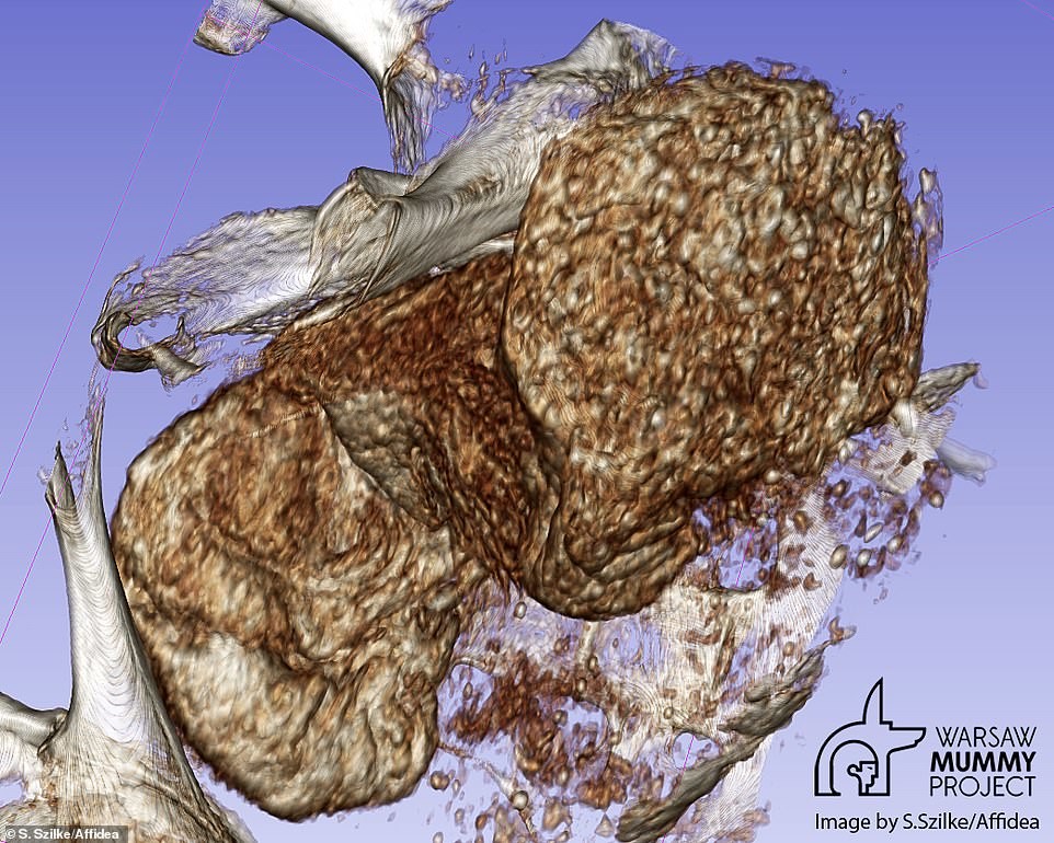
According to the position of the fetus and how the birth canal closed, researchers were able to determine that the "mysterious lady" died without childbirth. In July, it was revealed that the mummified woman was most likely died of nasopharyngeal cancer, a rare cancer that affects part of the throat connects the back of the nose to the back of the mouth. Researchers The Warsaw Mummy Project scanned the skull of an ancient corpse, when they found unusual marks on the bone, indicating cancer. 